
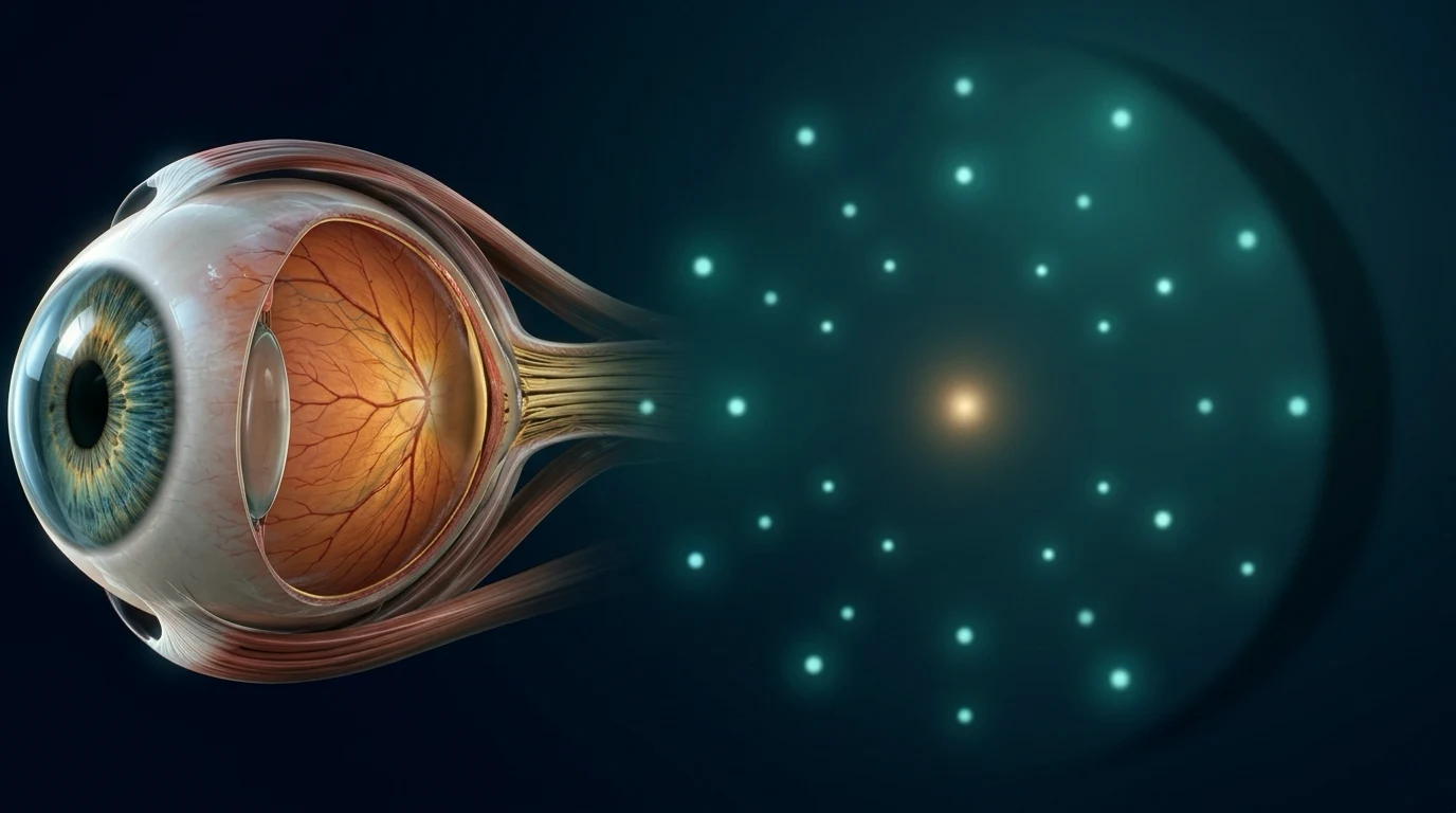
فحص مجال النظر، أو قياس المجال البصري، يقيس ما تستطيع العين رؤيته حول نقطة ثابتة أمامك، وليس حدة النظر على اللوحة فقط. أثناء الفحص تنظر إلى نقطة في جهاز وتضغط زرًا عندما ترى ومضة خفيفة في أماكن مختلفة. يُستخدم كثيرًا في متابعة الجلوكوما وتقييم العصب البصري وبعض مشاكل المسارات البصرية. الفحص غير مؤلم ولا يستخدم أشعة، لكن النتيجة تحتاج تركيزًا وتفسيرًا مع ضغط العين وفحص العصب وOCT. نقطة حمراء واحدة أو فحص متعب لا تشخص مرضًا بمفردها، وقد يطلب الطبيب إعادة الفحص للتأكد من النمط.
ماذا يقيس فحص مجال النظر؟
حدة النظر تخبرنا بقدرتك على رؤية تفاصيل أمامك مباشرة، مثل الحروف على اللوحة. أما مجال النظر فيبحث عن مناطق قد تكون أضعف أو مفقودة حول نقطة التثبيت، بما فيها الرؤية الجانبية وأجزاء من المجال القريب من المركز. العينان قد تتعاونان في الحياة اليومية، لذلك يفحص الجهاز كل عين على حدة لمعرفة الصورة بدقة.
الفحص الأكثر شيوعًا هو فحص آلي ثابت: تظهر ومضات بدرجات سطوع مختلفة، ويقيس الجهاز أضعف ومضة لاحظتها في كل نقطة. هذا لا يعني أن عليك أن تبحث بعينك عن الومضات، بل أن تثبت النظر في المنتصف وتستجيب فقط عندما تراها من دون تحريك العين.
ماذا يحدث أثناء الفحص؟
تجلس أمام جهاز على شكل قبة، وتغطي عينًا بينما تبقى العين الأخرى مثبتة على نقطة في الوسط. عندما ترى ومضة، اضغط الزر مرة واحدة. بعض الومضات خافتة جدًا، لذلك لا تخمّن ولا تضغط إذا لم تر شيئًا. إذا احتجت أن ترمش أو ترتاح لثوانٍ، أخبر الفني بدل أن تكمل وأنت متوتر.
قد يستخدم المركز عدسة تجريبية داخل الجهاز لتصحيح المسافة القريبة. مدة الفحص تختلف حسب البرنامج وقد تطول قليلًا في أول مرة. التعلّم الطبيعي والقلق أو التعب قد يؤثرون في الأداء، ولذلك لا تعتبر النتيجة الأولى حكمًا نهائيًا على العين.
لماذا يطلبه الطبيب؟
في الجلوكوما، يساعد فحص مجال النظر على معرفة هل تأثرت وظيفة العصب البصري وهل يوجد تغير مع الوقت. لا يكفي قياس ضغط العين وحده، لأن بعض الناس لديهم ضغط ضمن الحدود المعتادة مع تغيرات في العصب أو المجال، بينما قد يكون ضغط العين مرتفعًا من دون ضرر واضح في الفحص الأول.
قد يُطلب الفحص أيضًا عندما تكون هناك مشكلة في العصب البصري، أو أعراض أو نتائج تشير إلى المسارات البصرية في المخ، وفي بعض أمراض الشبكية. نوع البرنامج وعدد مرات التكرار يحددهما السؤال الطبي، لذلك لا تقارن نتيجتك مباشرة بنتيجة شخص آخر أو ببرنامج مختلف.
هل يغني عن OCT أو فحص العصب؟
لا. فحص OCT يصور طبقات العصب أو الشبكية، بينما مجال النظر يقيس وظيفة الرؤية. قد تظهر تغيرات في أحدهما قبل الآخر، وقد تتأثر النتيجة بجودة الصورة أو التركيز أو درجة النظارة. لهذا يربط الطبيب بين الأعراض، الضغط، شكل العصب، صور OCT، وفحوص المجال المتتابعة بدل الاعتماد على اختبار واحد.
الورقة المطبوعة فيها ألوان وأرقام وخريطة رمادية، لكن أهم شيء هو النمط وهل يتكرر وهل يتناسب مع بقية الفحص. وجود لون أحمر أو نقطة ناقصة لا يعني تلقائيًا جلوكوما أو فقدان نظر، كما أن الورقة الطبيعية وحدها لا تنفي كل مشكلة في مرحلة مبكرة.
كيف تستعد للحصول على نتيجة مفيدة؟
نم جيدًا قدر الإمكان، وخذ نظارتك الحالية وأبلغ الفريق إذا تغير مقاسها أو كانت الرؤية ضبابية. استخدم أدويتك المعتادة ما لم يطلب الطبيب غير ذلك. إذا كنت تعاني من جفاف أو حساسية شديدة في العين، أخبرهم قبل البدء لأن الراحة والترطيب المناسبين قد يساعدانك على تثبيت النظر بشكل أفضل.
لا تقلق من ضياع ومضة أو اثنتين. المطلوب أن تكون صادقًا وهادئًا، لا أن تحصل على نتيجة مثالية. لا تغير طريقة الضغط من نفسك ولا تنظر إلى جوانب القبة. لو شعرت بالتعب، اطلب استراحة قصيرة. وقد يعاد الاختبار في زيارة أخرى بدل اتخاذ قرار كبير من نتيجة غير موثوقة.
متى لا أنتظر موعد فحص المجال؟
فحص المجال متابعة مهمة، لكنه ليس بديلًا عن التقييم العاجل. فقدان مفاجئ أو ستارة في جزء من النظر، ومضات جديدة كثيرة مع عوامات، ألم شديد أو احمرار شديد، أو صداع جديد مع ازدواج رؤية أو أعراض عصبية يحتاج تواصلًا عاجلًا مع طبيب العيون أو الطوارئ حسب الحالة.
لو كان لديك جلوكوما، لا توقف القطرات أو تغير الجرعة بسبب ورقة فحص واحدة. اسأل الطبيب عن النتيجة في سياق قياساتك السابقة والخطة التي تحمي العصب على المدى الطويل.
أسئلة شائعة
هل فحص مجال النظر مؤلم أو فيه أشعة؟
لا. هو اختبار ضوئي لا يلمس العين ولا يستخدم أشعة. قد يكون مرهقًا قليلًا لأنه يحتاج تركيزًا، لكن يمكن أخذ استراحة قصيرة.
هل أحتاج لتوسيع حدقة العين قبل الفحص؟
غالبًا لا يحتاج فحص المجال نفسه إلى توسيع. قد تُستخدم قطرات في الزيارة لأسباب أخرى، والطبيب أو الفريق سيخبرك بالترتيب المناسب.
هل نتيجة غير طبيعية تعني أن لدي جلوكوما؟
ليس بالضرورة. تحتاج النتيجة إلى مراجعة جودة الفحص وتكرار النمط وربطه بالضغط وشكل العصب وOCT وباقي الفحص.
كم مرة أحتاج الفحص؟
لا توجد إجابة واحدة. التكرار يعتمد على سبب الفحص، عوامل الخطورة، النتائج السابقة، وهل يوجد تغير يحتاج تأكيدًا.
تنبيه مهم: هذا المحتوى للتوعية ولا يغني عن الكشف عند طبيب العيون. إذا حدث فقدان مفاجئ في النظر، أو ألم شديد، أو ومضات جديدة، احجز فحصًا عاجلًا.
English version
Visual Field Testing: What It Measures and Why an Eye Specialist Orders It
What does a visual field test measure?
Visual acuity tells us how well you see fine detail straight ahead, such as letters on a chart. A visual field test looks for areas that may be weaker or missing around a fixation point, including peripheral vision and parts of the field near the center. Because both eyes work together in daily life, the machine tests one eye at a time to make the result specific.
The most common version is automated static perimetry: faint lights of different brightness appear while the machine estimates the dimmest light noticed at each location. You should not search for lights with your eyes. Keep looking at the center and respond only when a light is seen without moving the eye.
What happens during the test?
You sit in front of a bowl-shaped machine and cover one eye while the other keeps looking at a central target. Press the button once when you see a light. Some lights are deliberately very dim, so do not guess or press when you have not seen one. If you need to blink or rest briefly, tell the technician instead of continuing while tense.
The clinic may place a trial lens in the machine to correct near focus. Test length varies by program and can feel longer the first time. Normal learning, anxiety and fatigue can affect performance, so a first test is not a final verdict on the eye.
Why might the doctor order it?
In glaucoma, visual field testing helps determine whether optic-nerve function is affected and whether it is changing over time. Eye pressure alone is not enough, because some people have pressure in the usual range with optic-nerve or field changes, while others have elevated pressure without clear damage on an initial test.
The test may also be used when an optic-nerve problem, symptoms or other findings raise concern about the visual pathways in the brain, and in selected retinal disorders. The program and repeat schedule depend on the medical question, so a result should not be compared directly with someone else's or with a different test pattern.
Does it replace OCT or optic-nerve examination?
No. OCT images the optic-nerve or retinal layers, whereas visual field testing measures visual function. Change can appear in one before the other, and either result can be influenced by image quality, concentration or refractive blur. The clinician combines symptoms, pressure, optic-nerve appearance, OCT and serial fields instead of relying on one test.
The printout contains colors, numbers and a gray map, but the important question is whether there is a repeatable pattern that matches the rest of the examination. A red area or a single missed point does not automatically mean glaucoma or vision loss, and a normal sheet alone does not exclude every early problem.
How can you prepare for a useful result?
Sleep as well as you can, bring your current glasses and tell the team if the prescription has changed or vision is blurred. Take usual medicines unless the clinician advises otherwise. If you have significant dry eye or irritation, mention it before testing because appropriate comfort measures may help you keep steady fixation.
Do not worry about missing one or two lights. The goal is an honest, calm response, not a perfect score. Do not change your button-pressing strategy or look around the bowl. If you become tired, ask for a brief pause. The clinician may repeat the test at another visit rather than make an important decision from an unreliable field.
When should you not wait for a scheduled field test?
Visual fields are valuable for follow-up, but they do not replace urgent assessment. Sudden loss or a curtain in vision, a sudden shower of flashes and floaters, severe pain or redness, or a new headache with double vision or neurological symptoms requires urgent contact with an eye specialist or emergency care as appropriate.
If you have glaucoma, do not stop drops or change the dose because of one printout. Ask the clinician to interpret the result alongside prior measurements and the long-term plan to protect the optic nerve.
Frequently asked questions
Is a visual field test painful or does it use radiation?
No. It is a light-based test that does not touch the eye and uses no radiation. It can feel tiring because it requires concentration, but a short break is possible.
Do my pupils need to be dilated before the test?
The field test itself usually does not require dilation. Drops may be used for another part of the visit, and the clinic will explain the appropriate order.
Does an abnormal result mean I have glaucoma?
Not necessarily. The clinician reviews test quality, whether the pattern repeats, and how it fits with pressure, optic-nerve appearance, OCT and the rest of the examination.
How often will I need the test?
There is no one schedule for everyone. Frequency depends on why it was ordered, risk factors, previous results and whether a possible change needs confirmation.
Medical note: This content is for education and does not replace an ophthalmic examination. Sudden vision loss, severe pain or new flashes require urgent assessment.
المراجع الطبية | Medical references
- National Institute for Health and Care Excellence. Glaucoma: diagnosis and management (NG81), visual-field recommendations.
- Phu J, Khuu SK, Nivison-Smith L, Kalloniatis M. Standard automated perimetry for glaucoma and diseases of the retina and visual pathways: current and future perspectives. Progress in Retinal and Eye Research. 2025;104:101307. DOI 10.1016/j.preteyeres.2024.101307. PMID 39413870.
- WuDunn D, et al. Central Visual Field Testing in Early Glaucoma: A Report by the American Academy of Ophthalmology. Ophthalmology. 2024;131:240-248. DOI 10.1016/j.ophtha.2023.10.008. PMID 38069944.
- Banc A, Kedar S. Interpretation of the Visual Field in Neuro-ophthalmic Disorders. Current Neurology and Neuroscience Reports. 2024;24:67-81. DOI 10.1007/s11910-024-01332-3. PMID 38289405.
- American Academy of Ophthalmology EyeWiki. Glaucoma Data Standards, visual-field quality and repeat confirmation.