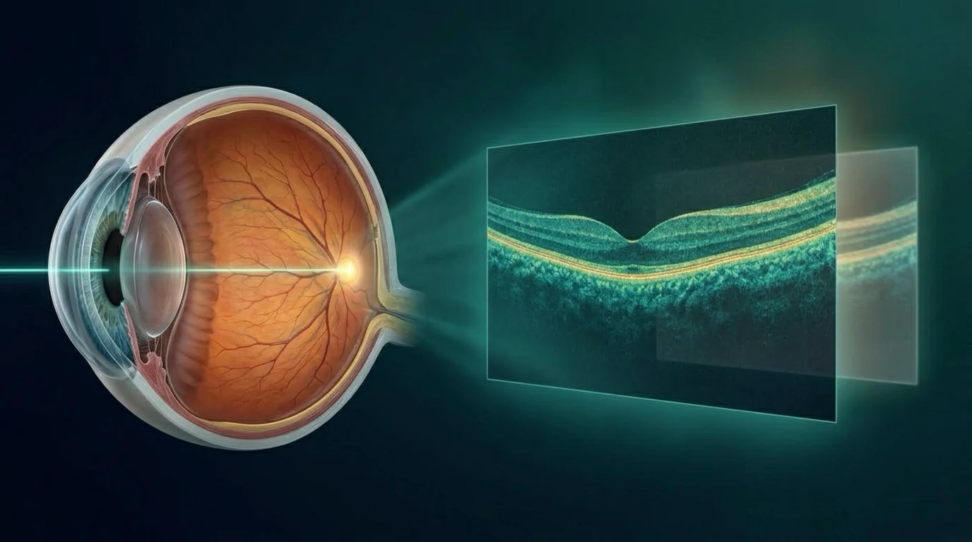
فحص OCT هو تصوير ضوئي غير مؤلم يعطي مقاطع دقيقة لطبقات الشبكية والعصب البصري، وأحيانًا القرنية. لا يستخدم أشعة سينية ولا يلمس العين، لكنه لا يغني عن كشف العين الكامل. قد يطلبه طبيب العيون لمتابعة الجلوكوما والسكري والتنكس البقعي وتورم الماكيولا أو مشاكل العصب، حسب الأعراض والكشف.
ماذا يرى جهاز OCT؟
التصوير يوضح سمك طبقات الشبكية، شكل الماكولا، وجود سوائل أو تورم، وحدود رأس العصب البصري. في OCT-A يمكن تقدير نمط الأوعية الدموية بدون صبغة في بعض الحالات.
في الجزء الأمامي يمكن استخدام أجهزة خاصة لتصوير القرنية وزاوية العين. نوع الفحص يختلف حسب السؤال الطبي، لذلك كلمة OCT وحدها لا تعني صورة واحدة لكل المرضى.
متى يطلبه الطبيب؟
في الجلوكوما يساعد OCT على قياس طبقة ألياف العصب البصري وخلايا العقدة ومقارنتها بالزيارات السابقة، لكنه لا يشخّص الجلوكوما وحده. في السكري والتنكس البقعي يكشف التورم أو السوائل ويساعد على متابعة الاستجابة للعلاج.
قد يُطلب عند ضعف نظر غير مفسر، أو تشوه الخطوط، أو الاشتباه في مشكلة بالعصب البصري. أما العوامات الجديدة فتحتاج أولًا إلى كشف سريع مع توسيع الحدقة لفحص الشبكية، وقد يضيف الطبيب OCT إذا احتاجه. اختيار التصوير مرتبط بالأعراض والكشف، وليس بمجرد طلب صورة للاطمئنان.
كيف يتم الفحص؟
تجلس أمام الجهاز وتثبت نظرك على علامة ضوئية لثوانٍ. لا يوجد تلامس ولا ألم، وقد يستخدم الطبيب قطرات توسّع حدقة العين إذا احتاج فحصًا أوسع أو كانت جودة الصورة ضعيفة.
النظارات أو العدسات لا تمنع الفحص عادة. أخبر الفريق إذا كان لديك صعوبة في تثبيت النظر أو إذا كنت تعاني من جفاف شديد، لأن الحركة والدموع قد تقللان جودة الصورة.
حدود الفحص وكيف تُقرأ النتيجة
قد تتأثر الأرقام بقصر النظر، عتامة العدسة، جفاف العين، اختلاف الجهاز أو جودة التثبيت. التقرير الآلي قد يضع علامة غير طبيعية تحتاج مراجعة، ولا يعني وحده وجود مرض أو تدهور.
الأهم هو مقارنة الصور الجيدة من نفس الجهاز مع الكشف على العين، لمعرفة هل التغيّر حقيقي أم مجرد اختلاف في القياس. لا تقارن رقمًا من الإنترنت برقمك من غير سياق.
هل OCT يغني عن الفحص؟
لا. الطبيب يربط الصورة بحدة النظر وضغط العين وشكل العصب وفحص القرنية والشبكية والأعراض. قد تكون صورة OCT طبيعية مع وجود مشكلة في جزء آخر من العين، وقد تكون غير مثالية لأسباب تقنية من غير وجود مرض.
إذا حدث فقدان مفاجئ للنظر أو ألم شديد أو ومضات وعوامات جديدة، لا تنتظر موعد OCT الروتيني. الأولوية للفحص العاجل، وقد يضاف التصوير بعده.
أسئلة شائعة
هل فحص OCT مؤلم؟
عادةً لا. هو تصوير من غير لمس العين، وقد تُستخدم قطرات توسّع حدقة العين حسب هدف الزيارة.
هل OCT يكتشف كل أمراض العين؟
لا. هو ممتاز لأسئلة معينة في الشبكية والعصب والقرنية، لكنه لا يغني عن الفحص ولا يلتقط كل الأمراض.
هل أحتاج OCT كل سنة؟
ليس بالضرورة. التكرار يعتمد على المرض وعوامل الخطورة وجودة الصور السابقة وقرار الطبيب.
هل الرقم الأحمر في التقرير يعني أنني سأفقد النظر؟
ليس وحده. الألوان قد تشير إلى مقارنة إحصائية أو جودة قياس، ويجب تفسيرها مع الطبيب والفحص والصور السابقة.
تنبيه مهم: هذا المحتوى للتوعية ولا يغني عن الكشف عند طبيب العيون. إذا حدث فقدان مفاجئ في النظر، أو ألم شديد، أو ومضات جديدة، احجز فحصًا عاجلًا.
English version
OCT Eye Scan: What Does It Show and When Is It Needed?
What does OCT show?
OCT can show retinal-layer thickness, macular contour, fluid or swelling and optic-nerve-head structure. OCT angiography can map aspects of retinal blood flow without dye in selected situations.
Specialized anterior-segment OCT can image the cornea and angle. The scan protocol depends on the clinical question, so an OCT is not one identical test for every patient.
When is OCT requested?
In glaucoma, OCT measures retinal nerve-fiber and ganglion-cell patterns for comparison over time, but it does not diagnose glaucoma by itself. In diabetes and AMD, it can reveal fluid and guide treatment monitoring.
It may be requested for unexplained blur, distorted lines, new floaters or suspected optic-nerve disease. The scan should answer a clinical question rather than replace an examination.
What happens during the scan?
You sit at the device and fixate on a target for a few seconds. The scan is non-contact and painless. Dilating drops may be used when a wider retinal examination or better image quality is needed.
Glasses and contact lenses usually do not prevent OCT. Tell the team if fixation is difficult or the eyes are very dry because motion and an unstable tear film can reduce image quality.
Limitations and interpretation
Readings can be affected by myopia, lens opacity, dry eye, device differences and fixation quality. An automated warning is a prompt for review, not a diagnosis or proof of progression.
The useful interpretation combines high-quality serial scans with the clinical examination. A number copied from the internet cannot be compared with yours without context.
Does OCT replace an examination?
No. The clinician combines the scan with visual acuity, pressure, optic-nerve and retinal findings, corneal assessment and symptoms. A normal OCT can coexist with disease elsewhere, while an imperfect scan may reflect artifact rather than pathology.
Sudden vision loss, severe pain or new flashes and floaters should not wait for a routine OCT appointment. Urgent examination comes first and imaging can be added.
Frequently asked questions
Is an OCT scan painful?
Usually not. It is a non-contact scan, although dilating drops may be used depending on the visit.
Does OCT detect every eye disease?
No. It is valuable for selected retinal, optic-nerve and corneal questions but does not replace a full examination.
Do I need OCT every year?
Not necessarily. Frequency depends on the condition, risk, previous image quality and the clinician's plan.
Does a red result mean I will lose vision?
Not by itself. Color coding may reflect a statistical comparison or artifact and must be interpreted with the examination and serial images.
Medical note: This content is for education and does not replace an ophthalmic examination. Sudden vision loss, severe pain or new flashes require urgent assessment.
المراجع الطبية | Medical references
- Optical coherence tomography retinal imaging: technological advances and clinical applications. Annals of Translational Medicine. 2025. DOI 10.21037/atm-24-211. PMID 40438521.
- Clinical applications of anterior-segment OCT: updated review. Diagnostics. 2024;14:122. DOI 10.3390/diagnostics14020122. PMID 38248000.
- OCT angiography in the diagnosis of ocular disease. Clinical and Experimental Optometry. 2024. DOI 10.1080/08164622.2024.2323603. PMID 38452795.
- American Academy of Ophthalmology EyeWiki. Optical Coherence Tomography.