
إيه اللي يخلي الطبيب يشك في العصب البصري؟
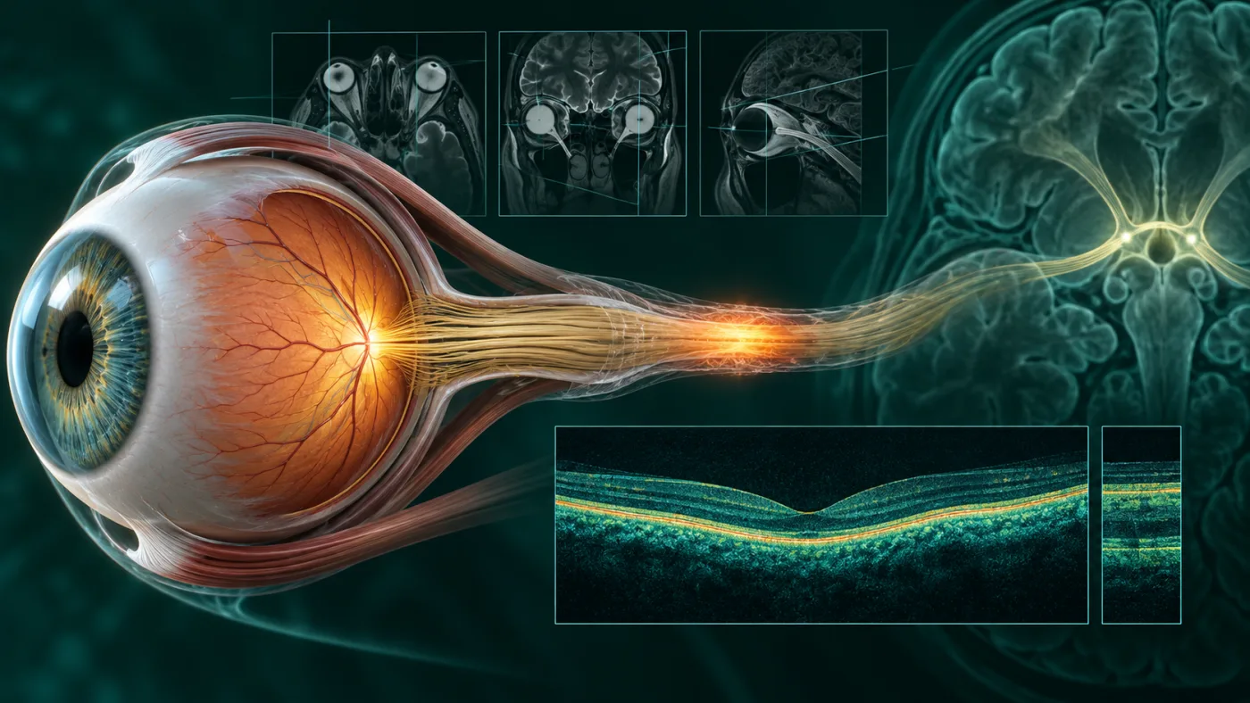
العصب البصري هو الكابل العصبي الذي ينقل المعلومات البصرية من العين إلى المخ. عندما يلتهب، قد يشعر المريض بألم خلف العين يزداد مع الحركة، ثم يلاحظ انخفاضًا في الرؤية أو أن الألوان - خصوصًا الأحمر - أصبحت باهتة. قد تتأثر الرؤية المركزية أو التباين حتى لو لم تكن العين حمراء من الخارج.
الأعراض غالبًا تتطور خلال ساعات إلى أيام. أي فقدان نظر جديد أو سريع يحتاج تقييمًا طبيًا سريعًا لأن أسباب ضعف النظر المفاجئ كثيرة وبعضها طارئ.
لماذا لم يعد مصطلح "التهاب العصب" كافيًا وحده؟
المراجعات الحديثة تؤكد أن optic neuritis له أسباب وأنماط متعددة. الشكل المرتبط بالتصلب المتعدد ليس هو نفسه التهاب العصب المرتبط بـneuromyelitis optica spectrum disorder أو MOG antibody-associated disease.
التمييز مهم لأن شدة النوبة، احتمال تكرارها، فرص استعادة الرؤية والحاجة لعلاج مناعي طويل المدى تختلف بين هذه الحالات. مراجعة 2025 في Continuum شددت على أهمية تعريف الأنماط بالأجسام المضادة بدل تجميعها كلها تحت "typical vs atypical" فقط.
دور MRI: أكثر من مجرد البحث عن التصلب المتعدد
MRI للمخ والمدارات مع بروتوكول مناسب يمكن أن يوضح موضع وطول الالتهاب في العصب، وجود enhancement، وأي آفات أخرى بالجهاز العصبي. بعض الأنماط التصويرية ترجح MS، وأخرى قد تكون أكثر توافقًا مع MOGAD أو NMOSD.
توقيت التصوير وجودته مهمان. لذلك طلب "MRI مخ" بشكل عام قد لا يساوي MRI موجهًا للعصب البصري والمدارات ببروتوكول مناسب.
ماذا يضيف OCT؟
Optical coherence tomography يقيس طبقات الشبكية المرتبطة بخلايا العصب البصري مثل retinal nerve fiber layer وganglion cell layer. في بداية النوبة قد توجد وذمة في بعض الحالات، وبعدها قد يظهر ترقق يعكس فقد ألياف عصبية.
OCT لا يشخص السبب وحده، لكنه أصبح أداة مهمة للمتابعة الكمية وربط ما يحدث في العصب بالوظيفة البصرية.
MOG-IgG وAQP4-IgG: تحاليل غيرت التصنيف
في الحالات التي تحمل سمات غير نمطية - مثل فقدان شديد، إصابة متكررة، تورم بارز في رأس العصب، إصابة ثنائية، أو نمط MRI معين - قد يطلب الطبيب تحاليل أجسام مضادة مثل aquaporin-4 IgG وMOG-IgG باستخدام الاختبارات المناسبة.
النتيجة قد تغير الخطة جذريًا؛ لأن NMOSD وMOGAD لهما استراتيجيات علاج ومنع تكرار مختلفة عن optic neuritis المرتبط بـMS.
هل الكورتيزون يعيد النظر؟
في optic neuritis الكلاسيكي المرتبط بالتصلب المتعدد، الكورتيزون الوريدي بجرعات عالية قد يسرع استعادة الرؤية، لكنه لا يعني بالضرورة تحسنًا أكبر في النتيجة النهائية طويلة المدى لكل حالة. أما في الأنماط المناعية الأخرى، توقيت العلاج وشدته قد يكونان أكثر أهمية، وقد تُستخدم plasma exchange في حالات شديدة لا تستجيب كما ينبغي.
وده سبب إضافي لعدم علاج "التهاب العصب" كتشخيص واحد ثابت. تحديد النوع والسياق العصبي/المناعي جزء من العلاج نفسه.
علامات تستحق تقييمًا سريعًا
ضعف رؤية سريع في عين واحدة أو العينين، ألم واضح مع حركة العين، انخفاض ملحوظ في رؤية الألوان، أو وجود أعراض عصبية أخرى يستحق تقييمًا عاجلًا لدى طبيب عيون/أعصاب عيون، وقد يتطلب تنسيقًا مع طبيب أعصاب.
أي فقدان مفاجئ شديد للرؤية، خصوصًا دون ألم أو مع أعراض عصبية حادة، يحتاج استبعاد أسباب أخرى طارئة مثل انسداد الأوعية أو أسباب عصبية مركزية.
الخلاصة
الـneuro-ophthalmology الحديثة تحولت من سؤال "هل هذا التهاب عصب؟" إلى "أي نوع من التهاب العصب، ولماذا؟". الجمع بين التاريخ المرضي، فحص العصب والرؤية اللونية، MRI، OCT والتحاليل الموجهة يسمح بخطة أدق وإنذار أوضح للمريض.
أسئلة شائعة
هل التهاب العصب البصري يعني بالضرورة تصلب متعدد؟
لا. MS سبب مهم، لكن توجد أسباب أخرى منها MOGAD وNMOSD وأسباب التهابية أو معدية أخرى.
هل ألم حركة العين علامة مهمة؟
نعم، هو شائع في optic neuritis الكلاسيكي، لكنه ليس موجودًا في كل الحالات ولا يكفي وحده للتشخيص.
هل OCT يغني عن MRI؟
لا. كل فحص يقدم معلومة مختلفة؛ MRI يقيّم العصب والمسارات العصبية، وOCT يقيس طبقات الشبكية المرتبطة بالعصب.
هل كل الحالات تحتاج تحليل MOG وAQP4؟
ليس بالضرورة. تُطلب التحاليل حسب العمر، شدة ونمط الإصابة، التكرار، MRI، وباقي العلامات السريرية.
ملاحظة طبية: هذا المحتوى للتثقيف ولا يُغني عن فحص طبيب العيون، خصوصًا عند وجود فقدان مفاجئ للرؤية أو ألم شديد أو أعراض طارئة.
English version
Optic Neuritis: Eye-Movement Pain, Color Vision Loss and What Has Changed in Modern Diagnosis
What makes a clinician suspect optic neuritis?
The optic nerve carries visual information from the eye to the brain. Inflammation can cause pain behind the eye that worsens with eye movement, followed by reduced visual acuity, contrast sensitivity or color saturation. Red may appear washed out, and central vision can be affected even when the eye looks externally normal.
Symptoms often evolve over hours to days. Any new or rapidly progressive visual loss deserves prompt assessment because acute vision loss has many possible causes, some of which are emergencies.
Why is "optic neuritis" no longer a complete diagnosis?
Modern reviews emphasize that optic neuritis is a syndrome with multiple distinct causes. Multiple-sclerosis-associated optic neuritis is not the same disease as aquaporin-4-positive neuromyelitis optica spectrum disorder or MOG antibody-associated disease.
The distinction matters because attack severity, recurrence risk, visual prognosis and need for long-term immunotherapy differ. A 2025 Continuum review highlighted the shift toward antibody-defined entities rather than grouping everything only as "typical" or "atypical" optic neuritis.
MRI is used for more than looking for multiple sclerosis
MRI of the brain and orbits using an appropriate optic-nerve protocol can show the location and length of inflammation, contrast enhancement and associated central nervous system lesions. Certain imaging patterns are more typical of MS, while others may suggest MOGAD or NMOSD.
Timing and protocol quality matter. A generic brain MRI is not always equivalent to dedicated orbital and optic-nerve imaging.
What does OCT add?
Optical coherence tomography can quantify retinal layers linked to optic-nerve axons and ganglion cells, including the retinal nerve fiber layer and ganglion-cell complex. Early swelling can occur in some presentations; later thinning can reflect axonal loss.
OCT does not identify the cause on its own, but it has become an important tool for objective structural follow-up and correlation with visual function.
MOG-IgG and AQP4-IgG changed disease classification
In patients with atypical features - such as profound visual loss, recurrent attacks, marked optic-disc edema, bilateral disease or a suggestive MRI pattern - clinicians may request aquaporin-4 IgG and MOG-IgG testing using appropriate assays.
The result can substantially change management because NMOSD and MOGAD require different acute and relapse-prevention strategies from conventional MS-associated optic neuritis.
Do steroids restore vision?
In classic MS-associated optic neuritis, high-dose intravenous corticosteroids can accelerate visual recovery, but they do not necessarily improve the ultimate long-term visual outcome in every case. In other immune-mediated forms, rapid treatment may be more important, and plasma exchange can be considered for severe attacks that respond inadequately to steroids.
This is another reason not to treat "optic neuritis" as one fixed diagnosis. Determining the subtype and systemic context is part of treatment.
When should the patient be assessed urgently?
Rapid visual loss in one or both eyes, pain with eye movement, a marked change in color perception or associated neurological symptoms warrants prompt ophthalmic or neuro-ophthalmic assessment, often coordinated with neurology.
Sudden profound visual loss - especially without pain or with acute neurological deficits - also requires urgent exclusion of vascular and central neurological causes.
Bottom line
Modern neuro-ophthalmology has moved from asking only "Is this optic neuritis?" to "Which optic neuritis syndrome is this, and what is causing it?" History, color-vision testing, optic-nerve examination, MRI, OCT and targeted antibody testing can produce a more accurate diagnosis, prognosis and treatment plan.
Frequently asked questions
Does optic neuritis automatically mean multiple sclerosis?
No. MS is an important cause, but MOGAD, NMOSD and other inflammatory or infectious disorders can also cause optic neuritis.
Is pain with eye movement an important clue?
Yes, it is common in classic optic neuritis, but it is not present in every case and is not diagnostic by itself.
Can OCT replace MRI?
No. They provide different information: MRI evaluates the optic nerve and central nervous system, while OCT quantifies retinal layers related to optic-nerve structure.
Does every patient need MOG and AQP4 antibody testing?
No. Testing is targeted according to age, severity, recurrence, MRI pattern and other clinical features.
Medical note: This content is for education and does not replace an examination by an ophthalmologist, particularly for sudden vision loss, severe pain or other urgent symptoms.
المراجع الطبية | Medical references
- Eggenberger E. Optic Neuritis. Continuum (Minneap Minn). 2025;31(2):407-435. PMID 40179402.
- Chaitanuwong P, Moss HE. Optic neuritis: a comprehensive review of current therapies and emerging treatment strategies. Front Neurol. 2025. PMID 40606143.
- Consensus statement for the diagnosis and management of optic neuritis. 2026. PMID 42535805.
- Diagnosing optic neuritis in the changing landscape of diagnostic criteria. PMID 41266242.