
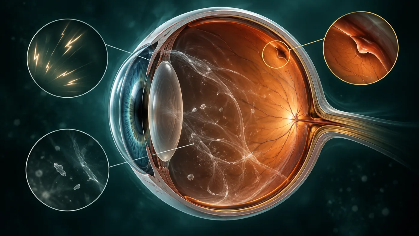
ما هي "الذبابة الطائرة" أصلًا؟
الأجسام الطافية أو floaters هي نقاط أو خيوط أو أشكال تتحرك مع النظر وتظهر أوضح أمام خلفية فاتحة. غالبًا تنتج من تغيرات طبيعية في الجسم الزجاجي، وهو الجل الشفاف الذي يملأ العين.
مع العمر قد ينكمش الجسم الزجاجي وينفصل عن سطح الشبكية فيما يسمى posterior vitreous detachment أو PVD. في كثير من الحالات يمر الأمر بدون مضاعفات، لكن في بعضها قد يجذب الجسم الزجاجي الشبكية بقوة ويسبب تمزقًا.
ليه الومضات مهمة؟
الومضات الضوئية قد تحدث عندما يحدث شد ميكانيكي على الشبكية. لذلك ظهور flashes مع floaters جديدة يرفع درجة الاهتمام، خصوصًا إذا ظهرت الأعراض فجأة في عين واحدة.
المراجعات العلمية تظهر أن وجود floaters وflashes معًا يرتبط بخطر أعلى لوجود retinal tear مقارنة ببعض الأعراض المنفردة. المهم ليس أن كل ومضة معناها انفصال، لكن لا يمكن تحديد السبب بأمان من الوصف وحده.
علامات الخطر التي لا تنتظر معها موعدًا عاديًا
اطلب فحصًا عاجلًا إذا ظهر فجأة عدد كبير من الـfloaters، أو ومضات جديدة متكررة، أو ظل/ستارة سوداء أو منطقة مفقودة من مجال الإبصار، أو انخفاض مفاجئ بالرؤية. الـNational Eye Institute يعتبر retinal detachment حالة طبية طارئة.
وجود نزيف داخل العين قد يظهر كدخان أو سحابة أو نقاط كثيرة، وقد يرتبط بتمزق في الشبكية أو بأسباب أخرى مثل اعتلال الشبكية السكري.
مين أكثر عرضة؟
الـNEI يذكر عوامل خطر تشمل قصر النظر الشديد، جراحة المياه البيضاء، إصابات العين، تاريخ سابق لانفصال الشبكية، وبعض أمراض الشبكية مثل اعتلال الشبكية السكري.
لكن غياب عوامل الخطر لا يستبعد المشكلة. أي تغير مفاجئ جديد يستحق التقييم حسب وصف الأعراض والفحص.
كيف يفحص الطبيب الشبكية؟
الفحص الأساسي هو dilated retinal examination بعد توسيع حدقة العين، وقد يستخدم الطبيب عدسات خاصة لفحص أطراف الشبكية. لو الرؤية الداخلية غير واضحة بسبب نزيف، قد يحتاج الأمر إلى ocular ultrasound في بعض الحالات.
لو وُجد retinal tear قبل حدوث انفصال واسع، قد يمكن علاجه بالليزر أو cryotherapy حسب الحالة لمنع وصول السائل خلف الشبكية. أما الانفصال نفسه فقد يحتاج جراحة مثل vitrectomy أو scleral buckle أو gas procedure وفق نوعه ومكانه.
الخلاصة
الذباب الطائر المزمن المستقر غالبًا أقل قلقًا من تغيير مفاجئ جديد. قاعدة الأمان سهلة: "floaters جديدة كثيرة + flashes، أو curtain/shadow = فحص عاجل". لأن أفضل وقت لعلاج تمزق الشبكية هو قبل ما يتحول لانفصال أكبر.
أسئلة شائعة
هل الذبابة الطائرة خطيرة دائمًا؟
لا. كثير منها حميد ومستقر، لكن ظهورها فجأة وبكثرة أو مع ومضات أو ستارة في النظر يحتاج فحصًا عاجلًا.
هل انفصال الجسم الزجاجي هو نفسه انفصال الشبكية؟
لا. PVD شائع مع العمر وغالبًا لا يسبب انفصال الشبكية، لكنه قد يسبب تمزقًا عند بعض المرضى.
هل يمكن أن يحدث انفصال شبكية بدون ألم؟
نعم، غالبًا لا يكون الألم هو العرض الأساسي. الأهم هو التغير المفاجئ في floaters أو flashes أو الظل بمجال الرؤية.
هل يكفي تصوير OCT؟
OCT ممتاز للماكولا والعصب، لكن تقييم تمزقات أطراف الشبكية يعتمد أساسًا على فحص قاع العين الموسع، وأحيانًا فحوص أخرى.
ملاحظة طبية: هذا المحتوى للتثقيف ولا يُغني عن فحص طبيب العيون، خصوصًا عند وجود فقدان مفاجئ للرؤية أو ألم شديد أو أعراض طارئة.
English version
Eye Floaters and Flashes: When Are They Benign and When Could They Signal Retinal Detachment?
What are floaters?
Floaters are moving dots, threads or cobweb-like shapes that are most visible against a bright background. They commonly arise from age-related changes in the vitreous gel that fills the eye.
As the vitreous liquefies and contracts, it can separate from the retina in a posterior vitreous detachment, or PVD. Most PVDs do not cause major complications, but vitreous traction can occasionally create a retinal tear.
Why do flashes matter?
Flashes can occur when the vitreous mechanically tugs on the retina. New flashes occurring together with sudden new floaters therefore deserve particular attention, especially when symptoms begin abruptly in one eye.
Systematic-review data show that combinations of floaters and flashes are associated with a higher probability of retinal tear than some isolated symptoms. This does not mean every flash equals retinal detachment; it means the cause cannot be safely determined from symptoms alone.
Warning signs that should not wait for a routine appointment
Seek urgent eye assessment for a sudden shower of floaters, repeated new flashes, a dark curtain or shadow, a missing area of peripheral vision or sudden loss of vision. The National Eye Institute classifies retinal detachment as a medical emergency.
Bleeding into the vitreous can appear as smoke, a cloud or many dark spots and may occur with retinal tears or other diseases such as diabetic retinopathy.
Who is at higher risk?
Risk factors described by the National Eye Institute include high myopia, prior cataract surgery, eye trauma, a personal or family history of retinal detachment and retinal disease such as diabetic retinopathy.
Absence of these factors does not rule out a tear. A sudden new symptom pattern still needs assessment based on urgency and examination findings.
How does the doctor check the retina?
The key examination is a dilated retinal examination using specialized lenses to inspect the peripheral retina. If blood or media opacity prevents a clear view, ocular ultrasonography may be needed.
A retinal tear found before extensive detachment can often be sealed with laser or cryotherapy in selected cases. Established retinal detachment may require procedures such as vitrectomy, scleral buckle or intraocular gas depending on the anatomy.
Bottom line
Long-standing stable floaters are generally less concerning than a sudden change. A useful safety rule is: "many new floaters plus flashes, or a curtain/shadow in vision, requires urgent retinal assessment." The best opportunity to protect vision is often to treat a retinal tear before a larger detachment develops.
Frequently asked questions
Are floaters always dangerous?
No. Many are benign and stable, but sudden numerous floaters or floaters with flashes or a curtain-like shadow require urgent examination.
Is posterior vitreous detachment the same as retinal detachment?
No. PVD is common with aging and often uncomplicated, but it can occasionally create a retinal tear.
Can retinal detachment happen without pain?
Yes. Pain is not a typical warning symptom. Sudden floaters, flashes, shadow or field loss are more important.
Is an OCT scan enough to rule out a retinal tear?
No. OCT is excellent for the macula and optic nerve, but peripheral retinal tears are primarily assessed with a dilated retinal examination and sometimes additional imaging.
Medical note: This content is for education and does not replace an examination by an ophthalmologist, particularly for sudden vision loss, severe pain or other urgent symptoms.