
المشكلة الأساسية: الشبكية قد تتأذى قبل ما تحس
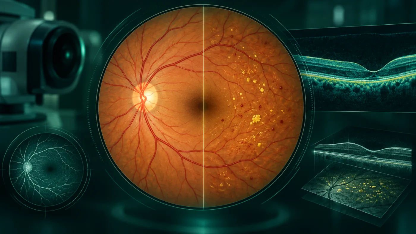
ارتفاع السكر المزمن يؤثر على الأوعية الدقيقة في الشبكية. في البداية قد تظهر microaneurysms وتسريب أو انسداد بأوعية صغيرة من غير ما المريض يحس بأي تغير. مع التقدم قد يحدث نقص تروية، أوعية دموية جديدة هشة، نزيف داخل العين، أو ارتشاح في مركز الإبصار.
الـNational Eye Institute يذكر بوضوح أن المراحل المبكرة غالبًا لا تسبب أعراضًا، ولذلك انتظار الزغللة قبل الفحص يفوّت أهم فرصة: اكتشاف التغيرات قبل فقدان النظر.
متى يبدأ فحص العين لمريض السكر؟
وفق Standards of Care in Diabetes 2026: مريض النوع الأول يحتاج فحصًا شاملًا موسعًا خلال خمس سنوات من بداية السكري. أما النوع الثاني، فالفحص يكون عند وقت التشخيص لأن المرض قد يكون موجودًا قبل اكتشافه بسنوات.
بعد ذلك يتحدد تكرار الفحص حسب وجود اعتلال بالشبكية، التحكم في السكر، والحالة العامة. لو لا يوجد اعتلال في فحوص متتالية وكان التحكم جيدًا، يمكن للطبيب في بعض الحالات إطالة الفاصل إلى كل سنة أو سنتين. أما وجود اعتلال فيحتاج متابعة سنوية على الأقل أو أقرب حسب الشدة.
ما هو ارتشاح مركز الإبصار DME؟
Diabetic macular edema يحدث عندما يتسرب السائل إلى منطقة الماكولا المسؤولة عن الرؤية المركزية الدقيقة. قد يسبب زغللة، تشوه خطوط أو صعوبة في القراءة، لكنه قد يُكتشف بالـOCT قبل شكوى شديدة.
الـADA 2026 تعتبر حقن anti-VEGF علاجًا أوليًا لمعظم العيون التي لديها center-involved DME مع ضعف في حدة الإبصار. الأدوية الحديثة وتوسيع فترات الجرعات عند بعض المرضى جعلت العلاج أكثر مرونة، لكن الانتظام في المتابعة عنصر أساسي.
متى نستخدم الليزر؟
الليزر ما زال مهمًا، خصوصًا panretinal photocoagulation في proliferative diabetic retinopathy عالية الخطورة، وفي بعض السيناريوهات الأخرى. كما يمكن أن يدخل focal/grid laser ضمن علاج بعض حالات الارتشاح المستمر أو عندما لا يكون anti-VEGF مناسبًا.
في proliferative retinopathy، anti-VEGF قد يكون بديلًا مناسبًا لليزر في مرضى مختارين، لكن يحتاج عادة التزامًا بزيارات وحقن متكررة. لو المريض معرّض للانقطاع عن المتابعة، تحمل الخطة العلاجية لهذه الحقيقة يصبح جزءًا من القرار.
السكر والضغط والدهون جزء من علاج العين
الـADA توصي بتحسين التحكم في السكر وضغط الدم والدهون لتقليل خطر حدوث أو تطور اعتلال الشبكية. علاج العين وحده لا يلغي أثر المرض العام.
لذلك أفضل رعاية لمريض الشبكية السكري تجمع بين طبيب العيون وطبيب السكر أو الباطنة، مع متابعة HbA1c والضغط والدهون بما يناسب حالة المريض العامة.
متى تصبح الحالة جراحية؟
لو حدث نزيف زجاجي شديد لا يتحسن، أو شد ليفي يهدد أو يفصل الشبكية، قد يحتاج المريض لعملية vitrectomy. القرار يعتمد على الرؤية، وضع الشبكية، كمية النزيف وشكل الشد.
الأهم أن كثيرًا من هذه المراحل المتقدمة يمكن تقليل خطر الوصول إليها لو اكتُشفت التغيرات وعولجت في التوقيت المناسب.
الخلاصة
فحص العين لمريض السكر ليس "فحص نظر" فقط، ولا يجب ربطه بوجود أعراض. هو فحص وقائي يحمي الرؤية قبل ما يشعر المريض أن هناك مشكلة. كلما كان التشخيص مبكرًا والـfollow-up منتظمًا، زادت الخيارات وقلت احتمالات فقدان رؤية لا يمكن استعادتها.
أسئلة شائعة
مريض السكر يفحص العين كل قد إيه؟
التوقيت يعتمد على نوع السكر ونتيجة الفحص. النوع الثاني يبدأ عند التشخيص، والنوع الأول خلال 5 سنوات من البداية، ثم يحدد الطبيب الفاصل حسب حالة الشبكية.
هل حقن العين مؤلمة؟
تُجرى عادة بتخدير موضعي وبتقنية معقمة، ويشعر كثير من المرضى بضغط أو انزعاج بسيط أكثر من ألم شديد.
هل الليزر أفضل من الحقن؟
ليس سؤال أفضل أو أسوأ. كل علاج له دور حسب وجود ارتشاح أو أوعية جديدة، مستوى الرؤية، والقدرة على الالتزام بالمتابعة.
هل ضبط السكر وحده يعالج اعتلال الشبكية؟
التحكم الجيد يقلل الخطر ويبطئ التقدم، لكنه لا يغني عن فحص وعلاج الشبكية إذا أصبحت التغيرات مهددة للنظر.
ملاحظة طبية: هذا المحتوى للتثقيف ولا يُغني عن فحص طبيب العيون، خصوصًا عند وجود فقدان مفاجئ للرؤية أو ألم شديد أو أعراض طارئة.
English version
Diabetic Retinopathy: Why You Should Not Wait for Vision Symptoms
The key problem: retinal damage can begin before you notice it
Chronic hyperglycemia damages the retinal microvasculature. Early changes can include microaneurysms, leakage and capillary closure without any subjective visual change. As disease advances, ischemia can drive fragile new vessels, vitreous hemorrhage or swelling in the macula.
The National Eye Institute emphasizes that early diabetic retinopathy often has no symptoms. Waiting for blur before arranging an eye examination therefore misses the main value of screening: finding disease before vision is threatened.
When should a person with diabetes start eye screening?
According to the ADA Standards of Care in Diabetes 2026, adults with type 1 diabetes should have an initial comprehensive dilated eye examination within five years after onset. People with type 2 diabetes should be examined at the time of diagnosis because hyperglycemia may have been present for years before diabetes was recognized.
Follow-up frequency then depends on retinal findings and systemic control. If repeated examinations show no retinopathy and glycemic indicators remain within target, intervals of one to two years may be considered in selected patients. Any retinopathy generally requires at least annual review, and more advanced disease needs closer surveillance.
What is diabetic macular edema?
Diabetic macular edema, or DME, occurs when fluid accumulates in the macula, the central retina responsible for detailed vision. It can cause central blur, distortion or reading difficulty, but OCT can detect structural change before severe symptoms develop.
ADA 2026 guidance identifies intravitreal anti-VEGF therapy as first-line treatment for most eyes with center-involved DME that is reducing visual acuity. Newer treatment regimens can sometimes extend intervals between injections, but reliable follow-up remains essential.
Where does laser still fit?
Retinal laser remains important, particularly panretinal photocoagulation for high-risk proliferative diabetic retinopathy and in selected other situations. Focal or grid laser can also have a role in persistent edema or when anti-VEGF therapy is unsuitable.
For proliferative retinopathy, anti-VEGF treatment can be an alternative to panretinal laser in selected patients, but it often requires repeated visits and injections. The patient's ability to maintain follow-up is therefore clinically important when choosing a strategy.
Glucose, blood pressure and lipids are part of eye treatment
The ADA recommends strategies to achieve individualized glycemic, blood-pressure and lipid goals because these measures can reduce the risk or slow progression of diabetic retinopathy. Eye injections or laser do not remove the effects of systemic disease.
The strongest care therefore combines retinal management with diabetes and cardiovascular risk management.
When does diabetic eye disease require surgery?
Vitrectomy may be needed for significant non-clearing vitreous hemorrhage, tractional retinal detachment or other advanced complications. The decision depends on the retina's anatomy, the level of vision and the degree of traction or bleeding.
Many advanced complications are more preventable when screening detects retinal disease early and treatment is delivered on schedule.
Bottom line
A diabetic eye examination is not merely a refraction check and should not depend on symptoms. It is preventive retinal care. Earlier detection and consistent follow-up preserve more options and reduce the risk of permanent vision loss.
Frequently asked questions
How often should a person with diabetes have an eye examination?
Timing depends on diabetes type and retinal findings. Type 2 diabetes should be screened at diagnosis; type 1 diabetes should receive an initial dilated exam within five years after onset, with follow-up individualized thereafter.
Are eye injections painful?
They are usually performed with topical anesthesia and sterile technique. Many patients describe pressure or brief discomfort rather than severe pain.
Is laser better than injections?
Neither is universally "better." The choice depends on whether the problem is macular edema, proliferative disease, visual acuity, anatomy and the ability to maintain follow-up.
Can good glucose control alone treat diabetic retinopathy?
Good systemic control reduces risk and slows progression, but it does not replace retinal treatment when sight-threatening disease is present.
Medical note: This content is for education and does not replace an examination by an ophthalmologist, particularly for sudden vision loss, severe pain or other urgent symptoms.