
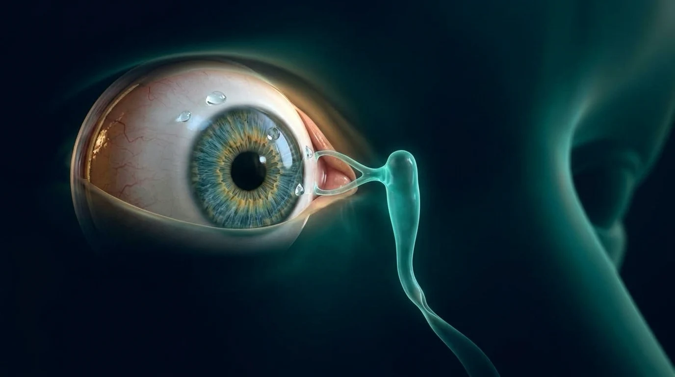
دموع مستمرة أو إفرازات لزجة من عين الرضيع قد تكون بسبب انسداد في القناة الدمعية، وليس بالضرورة التهابًا في العين. هذا الأمر شائع في الشهور الأولى، وكثير من الحالات تتحسن مع النمو والمتابعة. فحص طبيب عيون الأطفال مهم لتأكيد السبب، وشرح طريقة تدليك بسيطة إن كانت مناسبة، وتحديد هل يكفي الانتظار أم نحتاج إلى تسليك القناة في وقت لاحق. لا تستخدم قطرات أو ضغطًا قويًا على زاوية العين من نفسك.
ما هو انسداد القناة الدمعية عند الرضع؟
الدموع لا تخرج من العين وحدها؛ لها فتحات صغيرة عند الجفن ثم تمر في قناة تصل إلى الأنف. عند بعض الأطفال يولد المسار بغشاء رفيع عند نهايته أو بضيق يمنع الدموع من النزول بسهولة. فتظل العين دامعة، وقد تتجمع إفرازات عند الرموش، خصوصًا بعد النوم.
يُسمى هذا غالبًا انسداد القناة الدمعية الخِلقي. لا يعني أن عين الطفل ضعيفة، ولا أن هناك خطأ من الأهل. لكن الدموع والإفرازات وحدها لا تكفي لتأكيد التشخيص، لأن أسبابًا أخرى قد تشبهه وتحتاج فحصًا مختلفًا.
ما العلامات التي يلاحظها الأهل؟
أشهر العلامات هي دموع تزيد من غير بكاء، بلل متكرر في الخد، أو إفرازات بيضاء أو صفراء خفيفة تجعل الرموش تلتصق. قد تكون عين واحدة أو العينان معًا. في الانسداد البسيط يظل بياض العين طبيعيًا في الغالب، مع أن الإفرازات قد تعود بعد تنظيفها.
التهاب الملتحمة قد يسبب احمرارًا أو عدوى تنتشر بين أفراد البيت، والحساسية غالبًا تسبب حكة في الأطفال الأكبر. كما توجد حالات أقل شيوعًا لا نريد أن نفوّتها، لذلك من المهم أن يفحص الطبيب القرنية وضغط العين وشكل مسار الدموع بدل الاعتماد على صورة أو وصف من الإنترنت.
كيف يتأكد الطبيب؟
يسأل الطبيب متى بدأت الدموع، وهل ظهرت منذ الأيام أو الأسابيع الأولى، ثم يفحص سطح العين، الجفون، فتحة القناة عند زاوية العين القريبة من الأنف، والقرنية. أحيانًا يكفي هذا الفحص الهادئ لتأكيد الصورة وشرح الخطة للأهل.
لو كانت الدموع شديدة جدًا، أو يوجد احمرار مستمر، أو حساسية واضحة من الضوء، أو كبر في حجم العين أو عتامة في القرنية، يحتاج الطفل تقييمًا أسرع لاستبعاد أسباب أخرى. هذه علامات لا تُفسر تلقائيًا بانسداد بسيط في القناة.
هل التدليك يساعد؟
قد يوصي طبيب عيون الأطفال بتدليك لطيف في المكان والطريقة المناسبين لبعض الحالات. الفكرة هي وضع ضغط قصير وموجّه قرب زاوية العين الداخلية ثم التحرك إلى أسفل على مسار القناة، وليس عصر العين نفسها. من الأفضل أن يريك الطبيب أو الممرضة الطريقة أولًا، لأن الضغط القوي أو العشوائي قد يهيّج الجلد ولا يفتح القناة.
اغسل يديك قبل لمس المنطقة، ونظّف الإفرازات بلطف بقطعة نظيفة وماء مناسب بعد استشارة الفريق. لا تبدأ مضادًا حيويًا أو أي قطرة من المنزل؛ القطرة قد تعالج إفرازات مصاحبة في مواقف محددة، لكنها لا تزيل الانسداد نفسه.
متى ننتظر ومتى نفكر في تسليك القناة؟
كثير من حالات الانسداد تتحسن خلال السنة الأولى من العمر، لذلك قد تكون المتابعة مع التدليك الموجّه هي الخطة المناسبة في البداية إذا كانت العين هادئة والطفل بخير. قرار الانتظار لا يعني إهمال المتابعة؛ راقب التغيرات واحضر في الموعد الذي يحدده الطبيب.
إذا استمر الانسداد بعد عمر مناسب، أو تكررت التهابات أو ظهرت مشكلة معقدة، قد يناقش الطبيب تسليك القناة. لا يوجد موعد واحد يناسب كل طفل؛ العمر، شدة الأعراض، شكل الانسداد، والفحص هي التي تحدد الوقت المناسب. لا تؤجل زيارة التقييم فقط لأن شخصًا آخر قال إن الحالة ستتحسن وحدها.
ما المقصود بتسليك القناة؟
التسليك إجراء يمرر فيه طبيب العيون أداة دقيقة جدًا عبر فتحة القناة ليعبر الغشاء أو الضيق الذي يمنع نزول الدموع. في الأطفال الأصغر قد يختلف مكان وطريقة الإجراء عن الطفل الأكبر، وقد يحتاج الطفل الأكبر إلى تخدير عام حتى يكون الإجراء آمنًا ودقيقًا.
بعض الحالات التي لا تتحسن من المحاولة الأولى أو تبدو أكثر تعقيدًا قد تحتاج أنبوبًا سيليكونيًا مؤقتًا أو توسيعًا بالبالون. هذه ليست خطوات مطلوبة لكل طفل، ولا تُختار من غير فحص. اسأل الطبيب عن سبب الاختيار، التخدير، المتابعة، وما العلامات التي تستدعي الاتصال بعد الإجراء.
متى أطلب مساعدة عاجلة؟
اطلب تقييمًا سريعًا إذا أصبحت المنطقة عند زاوية العين الداخلية حمراء ومتورمة ومؤلمة، أو ظهرت حرارة أو خمول على الطفل، أو كان هناك تورم أزرق قرب الأنف. كذلك لا تنتظر إذا كانت العين حمراء جدًا، أو الطفل لا يحتمل الضوء، أو تبدو القرنية غائمة، أو تغيّر شكل العين بشكل واضح.
هذه العلامات لا تعني دائمًا مشكلة خطيرة، لكنها تحتاج فحصًا في الوقت المناسب لأن التهاب كيس الدمع أو سببًا آخر قد يحتاج علاجًا مختلفًا عن التدليك والمتابعة العادية.
أسئلة شائعة
هل الإفرازات تعني أن طفلي عنده التهاب دائمًا؟
لا. الإفرازات قد تظهر لأن الدموع لا تُصرف جيدًا، وقد تعود بعد تنظيفها. الاحمرار الشديد أو الألم أو تورم زاوية العين أو حرارة الطفل يحتاج تقييمًا سريعًا.
هل قطرة المضاد الحيوي تفتح القناة الدمعية؟
لا. قد يصف الطبيب قطرة في حالات محددة إذا كانت هناك إفرازات مصاحبة، لكنها لا تزيل الغشاء أو الضيق الذي يسبب الانسداد.
هل أعمل التدليك من غير شرح؟
الأفضل لا. المكان والاتجاه والضغط يهمون، ولذلك اطلب من طبيب عيون الأطفال أو الممرضة أن يريك الطريقة المناسبة لطفلك.
هل كل طفل يحتاج تسليكًا؟
لا. كثير من الحالات تتحسن مع النمو والمتابعة. يُناقش التسليك إذا استمر الانسداد أو ظهرت التهابات متكررة أو دلّ الفحص على حاجة لذلك.
تنبيه مهم: هذا المحتوى للتوعية ولا يغني عن الكشف عند طبيب العيون. إذا حدث فقدان مفاجئ في النظر، أو ألم شديد، أو ومضات جديدة، احجز فحصًا عاجلًا.
English version
Blocked Tear Duct in Babies: When Massage Is Enough and When Probing Is Considered
What is a blocked tear duct in a baby?
Tears do not simply leave the surface of the eye; they pass through tiny openings in the eyelids and then along a drainage pathway to the nose. In some babies, a thin membrane near the end of that pathway or a narrow segment keeps tears from draining easily. The eye may stay watery and lashes can collect discharge, especially after sleep.
This is commonly called congenital nasolacrimal duct obstruction. It does not mean that the child's eye is weak or that a parent did anything wrong. Tearing and discharge alone do not prove the diagnosis, though, because other problems can look similar and need a different examination.
What might parents notice?
The common signs are tears without crying, a cheek that stays wet or mild white-to-yellow discharge that makes the lashes stick together. It can affect one eye or both. With uncomplicated blockage, the white of the eye is often not very red, although discharge can return after it is cleaned away.
Conjunctivitis can cause more obvious redness or spread among people at home, while allergy more often causes itch in older children. Less common conditions should not be missed, so an eye specialist needs to examine the cornea, eye pressure and tear-drainage area rather than relying on a photograph or an online description.
How is it confirmed?
The eye specialist asks when tearing began and whether it was present in the first days or weeks, then examines the eye surface, eyelids, the area near the inner corner and the cornea. Often, this calm examination is enough to confirm the likely pattern and explain the plan.
Very heavy tearing, persistent redness, marked light sensitivity, an eye that appears larger or a cloudy cornea call for faster assessment to rule out other causes. These signs should not automatically be explained as a simple blocked duct.
Can massage help?
A pediatric eye specialist may recommend gentle massage for selected babies. The aim is a short, directed movement near the inner corner and downward along the drainage pathway, not squeezing the eyeball itself. It is best for a doctor or nurse to demonstrate the technique first, because forceful or random pressure can irritate the skin and does not open the duct.
Wash your hands before touching the area, and gently clean discharge with a clean material and an approach advised by the care team. Do not start antibiotic drops or other eye drops at home. A drop may be used for associated discharge in specific situations, but it does not remove the blockage itself.
When is observation enough, and when is probing discussed?
Many blocked ducts improve during the first year of life, so observation with guided massage may be the right initial plan when the eye is calm and the baby is otherwise well. Observation does not mean ignoring follow-up; watch for change and attend the review recommended by the specialist.
If blockage persists past an appropriate age, infections keep returning or the anatomy seems more complex, the specialist may discuss probing. There is no single deadline for every child: age, symptoms, the suspected type of blockage and examination findings shape the timing. Do not delay an assessment only because someone else's child improved without a procedure.
What does tear-duct probing involve?
Probing is a procedure in which an eye specialist passes a very fine instrument through the tear opening to pass the membrane or narrowing that is blocking drainage. The setting and approach may differ for younger and older children, and an older child may need general anesthesia so the procedure can be performed safely and accurately.
Some ducts that do not improve after an initial attempt or appear more complex may need temporary silicone intubation or balloon dilation. These are not needed for every child and should not be chosen without examination. Ask why an option is being suggested, what anesthesia and follow-up involve, and which signs should prompt a call after the procedure.
When should parents seek urgent help?
Seek prompt assessment if the inner corner becomes red, swollen and painful; if the baby has fever or seems unwell; or if there is a bluish swelling near the nose. Do not wait if the eye is very red, the child cannot tolerate light, the cornea looks cloudy or the eye's appearance changes noticeably.
These signs do not always mean a serious problem, but they deserve timely examination because an infected tear sac or another cause may need treatment that differs from ordinary massage and follow-up.
Frequently asked questions
Does discharge always mean my baby has an infection?
No. Discharge can happen because tears are not draining well and may return after cleaning. Marked redness, pain, swelling at the inner corner or fever need prompt assessment.
Do antibiotic drops open a blocked tear duct?
No. A doctor may prescribe drops in selected situations for associated discharge, but drops do not remove the membrane or narrowing that causes the blockage.
Can I start massage without being shown how?
It is better not to. The location, direction and pressure matter, so ask a pediatric eye specialist or nurse to show you the appropriate technique for your child.
Does every child need probing?
No. Many cases improve with growth and follow-up. Probing is discussed when blockage persists, infections recur or the examination shows that it is needed.
Medical note: This content is for education and does not replace an ophthalmic examination. Sudden vision loss, severe pain or new flashes require urgent assessment.
المراجع الطبية | Medical references
- American Association for Pediatric Ophthalmology and Strabismus. Nasolacrimal Duct Obstruction. Updated 2025.
- Sasaki T, et al. Congenital nasolacrimal duct obstruction: clinical guideline. Japanese Journal of Ophthalmology. 2024;68:367-388. DOI 10.1007/s10384-024-01064-4. PMID 39150609.
- Eshraghi B, et al. Assessing the success rate of treatment in simple and complex congenital nasolacrimal duct obstruction: a systematic review and meta-analysis. Graefe's Archive for Clinical and Experimental Ophthalmology. 2024;262:1993-2004. DOI 10.1007/s00417-023-06318-4. PMID 38059998.
- Alruwaili R, et al. Comparative Analysis of Silicone Tube Intubation Versus Probing and Balloon Dilation for Congenital Nasolacrimal Duct Obstruction: A Systematic Review and Meta-Analysis. Journal of Craniofacial Surgery. 2024;35:1114-1119. DOI 10.1097/SCS.0000000000010273. PMID 38727216.
- American Academy of Ophthalmology. Office- or Facility-Based Probing for Congenital Nasolacrimal Duct Obstruction: A Report. Ophthalmology. 2021. PMID 33358412.