
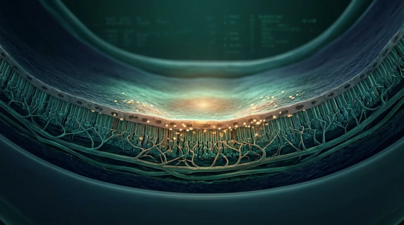
التنكس البقعي المرتبط بالعمر يؤثر في مركز النظر، وغالبًا لا يسبب ألمًا وقد يبدأ بأعراض خفيفة أو من غير أعراض. الكشف على قاع العين بعد توسيع الحدقة وتصوير OCT يساعدان الطبيب على معرفة إن كان التنكس جافًا، وهل وصل لمرحلة الضمور الجغرافي، أو تحوّل إلى النوع الرطب الذي قد يحتاج حقنًا داخل العين. لو لاحظت أن الخطوط المستقيمة بقت متعرجة أو ظهرت بقعة جديدة في مركز النظر، احجز كشفًا سريعًا.
ما هو التنكس البقعي؟
البقعة هي المنطقة المركزية من الشبكية التي نستخدمها للقراءة، والتعرف على الوجوه، ورؤية التفاصيل. في التنكس البقعي المرتبط بالعمر تتأثر هذه المنطقة تدريجيًا، بينما يظل النظر الطرفي موجودًا غالبًا.
قد تظهر في البداية ترسبات صفراء صغيرة تُسمى الدروزن (drusen). وجودها لا يعني أن فقدانًا شديدًا في النظر سيحدث حتمًا، لكنه سبب كافٍ للمتابعة والاهتمام بعوامل الخطورة.
علامات ممكن تظهر في البيت
الأعراض قد تشمل زغللة في مركز النظر، صعوبة القراءة رغم النظارة، بهتان الألوان، أو الحاجة لإضاءة أقوى. أحيانًا يلاحظ المريض أن الخطوط المستقيمة تبدو متموجة أو أن جزءًا من الكلمة يختفي.
جرّب تغطي عينًا وتقرأ أو تنظر إلى شبكة أمسلر إذا نصحك الطبيب بها، ثم كرر ذلك بالعين الثانية. الاختبار في البيت لا يشخّص المرض، لكنه قد يلفت انتباهك لتغيّر يحتاج كشفًا.
كيف يعرف الطبيب النوع والمرحلة؟
يبدأ الكشف بتوسيع الحدقة وفحص قاع العين، ثم تصوير OCT الذي يوضح طبقات الماكيولا بدقة. لو احتاج الطبيب تفاصيل أكثر، قد يطلب تصوير الأوعية بالفلوريسئين أو OCT-A لمعرفة هل توجد أوعية غير طبيعية أو تسريب.
المهم ليس اسم الحالة فقط، بل مرحلتها وهل فيها نشاط يحتاج علاجًا سريعًا. لذلك لا تكفي قراءة لوحة النظر وحدها لاتخاذ القرار.
الجاف، والضمور الجغرافي، والرطب
النوع الجاف هو الأكثر شيوعًا، وقد يظل مستقرًا فترة طويلة أو يتقدم تدريجيًا. في الضمور الجغرافي تحدث مناطق فقد في خلايا الشبكية، ويُناقش العلاج المتخصص حسب المكان والمرحلة والمخاطر.
في النوع الرطب تتكون أوعية دموية غير طبيعية تحت الشبكية وقد يحدث نزيف أو تورم. في حالات كثيرة يكون العلاج حقنًا داخل العين تقلل نشاط هذه الأوعية (Anti-VEGF)، مع متابعة منتظمة. طبيب الشبكية يختار الدواء ومواعيد الحقن حسب استجابة كل عين.
العلاج والحفاظ على الرؤية
في مراحل معينة من النوع الجاف قد يقترح الطبيب مكملات AREDS2. ليست ضرورية لكل مريض، فلا تبدأها من نفسك قبل معرفة المرحلة. لو كنت مدخنًا أو كنت تدخن سابقًا، أخبر الطبيب لتختار التركيبة المناسبة؛ التركيبات القديمة التي تحتوي على بيتا كاروتين لا تناسب المدخنين.
الامتناع عن التدخين، ضبط ضغط الدم والسكري، الحركة، وتناول غذاء متوازن غني بالخضروات كلها خطوات مفيدة للصحة العامة وصحة الشبكية. ارتدِ نظارة شمسية تحجب الأشعة فوق البنفسجية، والتزم بموعد المتابعة حتى لو كانت الأعراض قليلة.
متى تكون الحالة عاجلة؟
ظهور تشوه جديد في الخطوط، أو بقعة سوداء أو رمادية في مركز النظر، أو انخفاض مفاجئ في الرؤية قد يعني نشاطًا في النوع الرطب ويحتاج كشفًا عاجلًا. لا تنتظر موعد المتابعة الروتيني ولا تقد السيارة إذا كانت الرؤية غير آمنة.
أسئلة شائعة
هل التنكس البقعي يسبب العمى الكامل؟
غالبًا يؤثر في الرؤية المركزية أكثر من الطرفية. شدة التأثير تختلف حسب النوع والمرحلة وسرعة العلاج، لذلك المتابعة المبكرة مهمة.
هل توجد قطرة تعالج التنكس البقعي؟
لا توجد قطرة منزلية تعيد خلايا الماكولا. النوع الرطب قد يحتاج حقنًا داخل العين، والنوع الجاف له متابعة وخيارات محددة حسب المرحلة.
هل كل مريض يحتاج مكملات AREDS2؟
لا. فائدتها ترتبط بمراحل معينة من AMD، وقد لا تناسب بعض الأشخاص. اسأل طبيب الشبكية قبل استخدامها.
هل يمكن أن يكون التنكس البقعي بدون أعراض؟
نعم، خاصة في المراحل المبكرة. فحص قاع العين والتصوير يساعدان على اكتشافه قبل ضعف الرؤية الواضح.
تنبيه مهم: هذا المحتوى للتوعية ولا يغني عن الكشف عند طبيب العيون. إذا حدث فقدان مفاجئ في النظر، أو ألم شديد، أو ومضات جديدة، احجز فحصًا عاجلًا.
English version
Age-Related Macular Degeneration: How Is It Detected and Managed?
What is AMD?
The macula is the central part of the retina used for reading, recognizing faces and seeing fine detail. In AMD, macular tissue gradually becomes dysfunctional while peripheral vision is often preserved.
Small deposits called drusen may appear in early disease. Drusen do not mean that severe vision loss is inevitable, but they make monitoring and risk assessment important.
Symptoms patients may notice
Symptoms can include central blur, difficulty reading despite updated glasses, faded colors or a need for brighter light. Straight lines may look wavy, or a small part of a word may seem missing.
Cover one eye at a time while reading or using an Amsler grid if your clinician recommends it. Home monitoring does not diagnose AMD, but a new change should trigger an eye examination.
How is AMD diagnosed?
Assessment starts with a dilated retinal examination. OCT provides cross-sectional images of the macula, while fluorescein angiography or OCT angiography may be used when abnormal vessels or leakage are suspected.
The key question is not only whether AMD is present, but which stage and phenotype is present and whether there is active disease requiring prompt treatment. Visual acuity alone is not enough for that decision.
Dry AMD, geographic atrophy and wet AMD
Dry AMD is the most common form and may remain stable or progress slowly. Geographic atrophy refers to areas where retinal cells are lost; specialist treatment is discussed according to lesion location, stage and risk profile.
Wet AMD involves abnormal blood vessels beneath the retina that can leak or bleed. Anti-VEGF injections are the main treatment for many active cases and require planned monitoring. The medication and interval are individualized by a retina specialist.
Treatment and protecting vision
For selected stages of dry AMD, a clinician may discuss an AREDS2 formulation. Supplements are not appropriate for everyone and can interact with smoking status or medical conditions, so do not start them without staging and medical advice.
Stopping smoking, controlling blood pressure and diabetes, staying active and eating a balanced diet rich in vegetables support general and retinal health. UV-blocking sunglasses and scheduled follow-up remain useful even when symptoms are mild.
When is it urgent?
New line distortion, a central gray or dark spot, or sudden central vision loss can indicate active neovascular disease and needs urgent assessment. Do not wait for a routine appointment or drive if vision is unsafe.
Frequently asked questions
Does AMD cause complete blindness?
It mainly affects central vision, while peripheral vision is often retained. The impact varies by type, stage and treatment response, which is why early monitoring matters.
Is there an eye drop that cures AMD?
No home eye drop restores damaged macular cells. Active wet AMD may need injections, while dry AMD is managed according to stage and risk.
Does every patient need AREDS2 supplements?
No. Benefit is linked to specific AMD stages and supplements may be unsuitable for some people. Discuss them with a retina clinician.
Can AMD have no symptoms?
Yes, particularly in early stages. Retinal examination and imaging can detect changes before obvious vision loss.
Medical note: This content is for education and does not replace an ophthalmic examination. Sudden vision loss, severe pain or new flashes require urgent assessment.
المراجع الطبية | Medical references
- National Eye Institute. Age-related macular degeneration.
- Avacincaptad pegol for geographic atrophy: two-year GATHER2 phase 3 results. Ophthalmology. 2026. DOI 10.1016/j.ophtha.2025.12.011. PMID 41407269.
- Complement C3 or C5 inhibition for geographic atrophy: living systematic review and meta-analysis. Survey of Ophthalmology. 2024. DOI 10.1016/j.survophthal.2023.11.008. PMID 38008405.
- Lutein, zeaxanthin and omega-3 for AMD: AREDS2 randomized clinical trial. JAMA. 2013. DOI 10.1001/jama.2013.4997. PMID 23644932.