
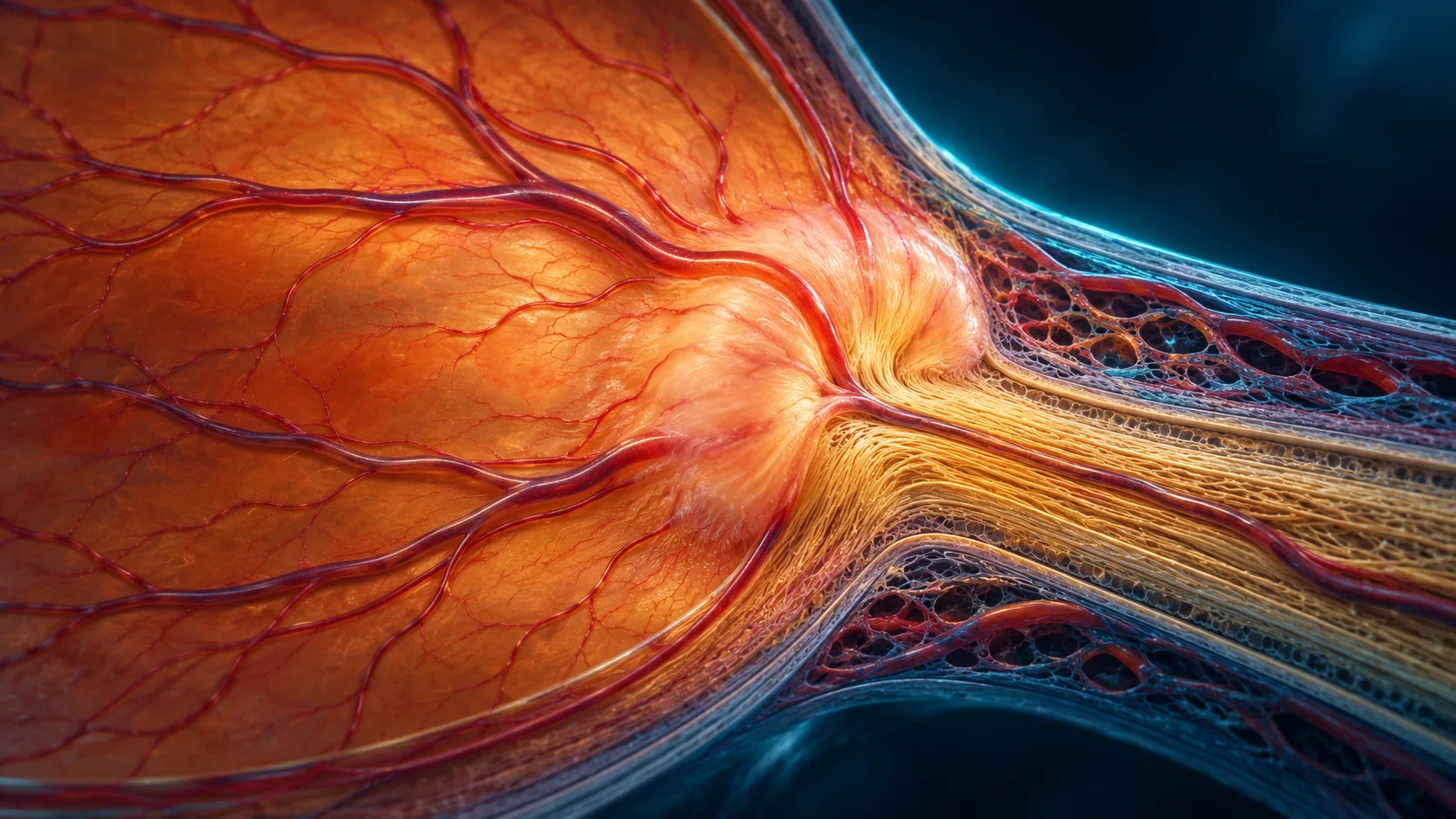
رأس العصب البصري هو المكان الذي يخرج منه العصب من العين إلى المخ. ظهوره متورمًا في الفحص له أسباب متعددة، وpapilledema اسم محدد للتورم المرتبط بارتفاع الضغط داخل الجمجمة. لا يمكن تحديد السبب من صورة واحدة أو الأعراض وحدها، وقد يحتاج الأمر تقييمًا عاجلًا.
هل كل تورم في العصب يعني ضغطًا على المخ؟
لا. قد يبدو العصب مرتفعًا بسبب اختلاف طبيعي أو ترسبات صغيرة أو أسباب أخرى. الطبيب يفرق بين التورم الحقيقي والمظهر الذي يشبهه باستخدام الفحص والصور والتاريخ الصحي، وأحيانًا فحوص إضافية.
ما الأعراض التي قد تصاحبه؟
قد لا يلاحظ الشخص أي تغير في البداية. أحيانًا تحدث نوبات قصيرة من تعتيم النظر أو صداع أو غثيان أو رؤية مزدوجة، لكن وجود هذه الأعراض أو غيابها لا يؤكد التشخيص. صداع شديد مفاجئ مع قيء أو ضعف عصبي يحتاج طوارئ.
كيف يتم التقييم؟
قد يشمل التقييم فحص نظر وحدقات ومجال النظر وتصوير OCT أو صورًا لقاع العين. إذا اشتبه الطبيب بسبب متعلق بالضغط داخل الرأس، قد ينسق تصويرًا للمخ وفحوصًا أخرى مع فريق الأعصاب أو الطوارئ. لا تؤخر التقييم عند الاشتباه.
لماذا العلاج يعتمد على السبب؟
تورم العصب وصف لعلامة في الفحص وليس تشخيصًا واحدًا. العلاج يختلف بحسب السبب، لذلك لا تبدأ علاجًا للضغط أو للعين من نفسك. احتفظ بنتائج الصور والفحوص واذكر للطبيب أي أدوية جديدة أو صداع أو تغير في النظر.
أسئلة شائعة
هل papilledema هو نفسه التهاب العصب البصري؟
لا. هما حالتان مختلفتان، ولكل منهما أسباب وفحوص وعلاج مختلف.
هل أحتاج إلى طوارئ إذا قال الطبيب إن العصب متورم؟
اتبع درجة الاستعجال التي يحددها الطبيب. عند صداع شديد مفاجئ أو قيء أو اضطراب عصبي، اذهب للطوارئ فورًا.
هل صورة قاع العين وحدها تكفي؟
قد تظهر التورم لكنها لا تحدد سببه دائمًا؛ قد يلزم فحص وتصوير أو اختبارات أخرى.
المقال للتوعية ولا يغني عن كشف طبيب العيون. فقدان النظر المفاجئ، أو الألم الشديد، أو ظهور ستار في مجال الرؤية يحتاج إلى تقييم عاجل.
English version
Optic Disc Swelling: When It May Signal Raised Pressure in the Head
Quick answer
The optic disc is where the optic nerve leaves the eye for the brain. A swollen appearance has several possible causes; papilledema specifically means swelling associated with raised pressure inside the skull. The cause cannot be determined from one image or symptoms alone, and urgent work-up may be needed.
Does every swollen-looking disc mean pressure in the head?
No. The disc may look elevated because of normal variation, small deposits or other causes. The ophthalmologist distinguishes true swelling from look-alikes using examination, imaging, health history and sometimes additional tests.
What symptoms can occur?
Some people notice no change at first. Brief episodes of dimmed vision, headache, nausea or double vision may occur, but these symptoms neither confirm nor exclude the diagnosis. A sudden severe headache with vomiting or neurological weakness needs emergency care.
How is it evaluated?
Assessment may include vision and pupil checks, visual fields, OCT or retinal photographs. If raised pressure in the skull is suspected, brain imaging and other tests may be coordinated with neurology or emergency teams. Do not delay evaluation when this is a concern.
Why does treatment depend on the cause?
Disc swelling is an examination finding, not a single diagnosis. Treatment differs by cause, so do not start eye or pressure medicines on your own. Bring imaging and test results and mention new medicines, headache or vision changes.
Frequently asked questions
Is papilledema the same as optic neuritis?
No. They are different conditions with different causes, tests and treatment.
Do I need emergency care if the disc is swollen?
Follow the urgency advised by your clinician. A sudden severe headache, vomiting or neurological symptoms needs emergency care now.
Is a fundus photo enough to diagnose the cause?
A photo may show swelling but does not always identify why; examination, imaging or other tests may be needed.
This article is for education and does not replace an eye examination. Sudden vision loss, severe pain or a curtain in your field of vision needs urgent assessment.