
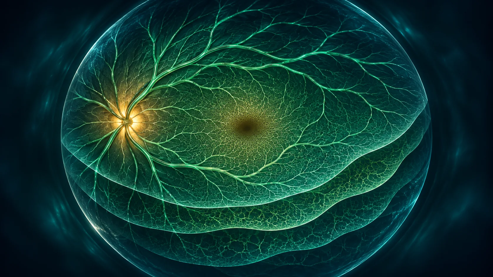
OCT angiography أو OCTA تصوير يلتقط حركة خلايا الدم داخل أوعية الشبكية باستخدام جهاز OCT، من دون حقن صبغة. يساعد في رسم بعض طبقات الأوعية، لكنه لا يجيب عن كل الأسئلة ولا يخلو من صور غير واضحة أو عيوب تقنية. يفسر الطبيب النتيجة مع فحص العين وباقي الصور.
كيف يتم تصوير OCTA؟
تضع ذقنك على الجهاز وتنظر إلى هدف ثابت بينما يلتقط الجهاز صورًا سريعة. لا تُحقن صبغة ولا يلمس الجهاز العين عادةً. قد تحتاج لإعادة التصوير إذا تحركت العين أو لم تكن الصورة واضحة، وقد تُستخدم قطرات توسيع حسب نوع الفحص.
ما الذي يمكن أن يوضحه؟
قد يعرض الجهاز تدفق الدم في طبقات مختلفة من الشبكية والمشيمية، ويساعد في تقييم بعض أمراض الأوعية أو مركز الإبصار. لا يمكن الاعتماد على صورة واحدة لتأكيد تشخيص أو تحديد علاج، لأن الطبيب يحتاج إلى الأعراض والفحص وقياسات أخرى.
ما الفرق بين OCT وOCTA؟
تصوير OCT العادي يوضح طبقات الشبكية وشكلها، أما OCTA فيضيف خريطة لحركة الدم في الأوعية. تصوير صبغة الشبكية يتتبع الصبغة داخل الأوعية وقد يظهر تسربًا بطريقة مختلفة. قد يحتاج الطبيب إلى أكثر من نوع تصوير.
حدود الفحص
حركة العين أو الرمش أو عتامة بعض أجزاء العين قد تؤثر في جودة الصورة. كما أن OCTA قد لا يظهر التسرب بنفس طريقة تصوير الصبغة. لذلك لا تقارن نتيجة بجهاز آخر أو تفسر الألوان بنفسك؛ اسأل الطبيب ما الذي تعنيه الصورة لحالتك.
أسئلة شائعة
هل OCTA يحتاج حقن صبغة؟
لا، التصوير نفسه لا يحتاج حقن صبغة، لكن قد يطلب الطبيب فحوصًا أخرى إذا احتاج معلومات إضافية.
هل OCTA يغني عن فحص قاع العين؟
لا. التصوير يكمل الفحص ولا يستبدل تقييم الطبيب للعين والنظر والأعراض.
هل OCTA مؤلم؟
عادةً لا؛ تنظر إلى هدف أمام الجهاز لثوانٍ أثناء التصوير.
المقال للتوعية ولا يغني عن كشف طبيب العيون. فقدان النظر المفاجئ أو الألم الشديد أو إصابة العين يحتاج إلى تقييم عاجل.
English version
OCT Angiography: Imaging Retinal Blood Flow Without Dye
Quick answer
OCT angiography, or OCTA, uses an OCT scanner to detect blood-cell motion in retinal vessels without injecting dye. It can map selected vessel layers, but it does not answer every question and may have image artifacts. The ophthalmologist interprets it alongside the eye exam and other imaging.
How is OCTA performed?
You rest your chin on the scanner and look at a target while it captures rapid images. No dye is injected, and the device usually does not touch the eye. Images may need repeating if the eye moves or the scan is unclear; dilation may be used depending on the test.
What can it show?
The scan can display blood flow in different retinal and choroidal layers and may help assess some vascular or macular conditions. One image cannot confirm a diagnosis or determine treatment by itself; symptoms, examination and other measurements matter.
OCT versus OCTA
Standard OCT shows retinal structure and layers; OCTA adds a map of blood flow in vessels. Dye angiography follows injected dye and can show leakage differently. An ophthalmologist may need more than one imaging method.
Limitations of the scan
Eye movement, blinking or media opacity can affect image quality. OCTA may not show leakage in the same way as dye angiography. Avoid comparing scans from different devices or interpreting colors alone; ask what the findings mean for your situation.
Frequently asked questions
Does OCTA require a dye injection?
No, the OCTA scan itself does not require injected dye, though other tests may be ordered for more information.
Does OCTA replace a retinal examination?
No. Imaging complements the eye exam and the clinician's assessment of vision and symptoms.
Is OCTA painful?
Usually not; you look at a target while the scanner captures images for a few seconds.
This article is for education and does not replace an eye examination. Sudden vision loss, severe pain or an eye injury needs urgent assessment.