
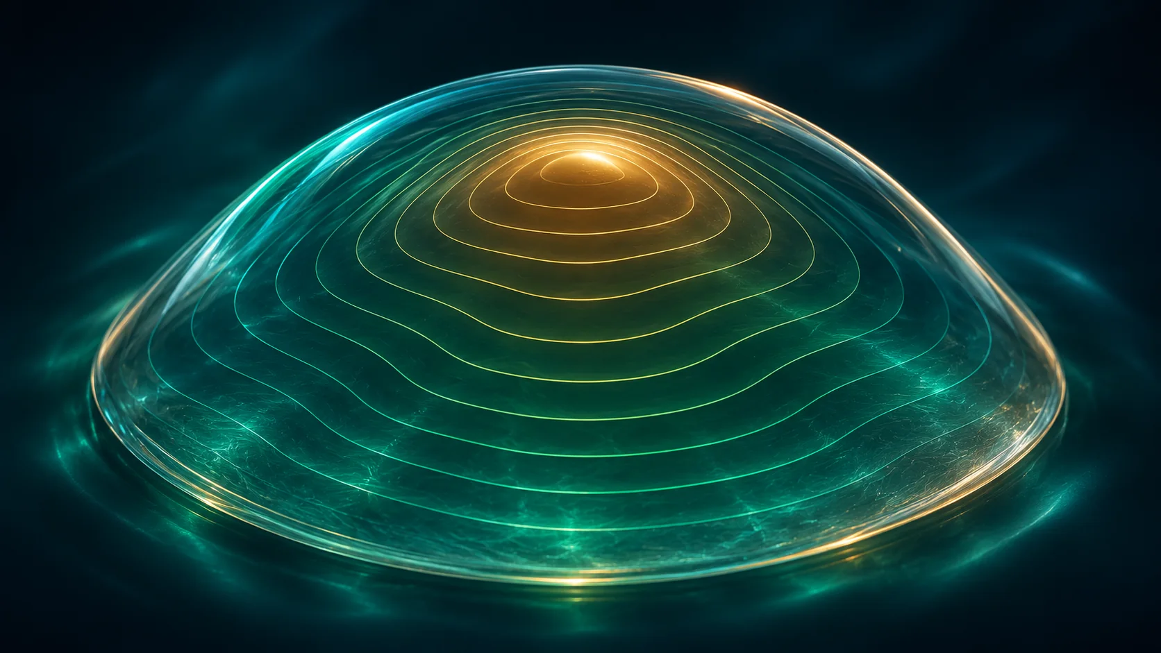
خريطة القرنية فحص تصويري يرسم شكل سطح القرنية وانحناءها، وبعض الأجهزة تقيس أيضًا سطحها الخلفي أو سماكتها. قد يطلبه الطبيب عند الاشتباه بالقرنية المخروطية أو قبل الليزك واختيار بعض العدسات. الفحص سريع ولا يلمس العين، لكن قراءة الخريطة تحتاج إلى طبيب يجمعها مع باقي القياسات.
إزاي يتم الفحص؟
تجلس أمام جهاز وتنظر إلى نقطة أو هدف ثابت لثوانٍ، بينما يلتقط الجهاز صورًا لسطح القرنية من دون لمس العين. غالبًا لا يحتاج إلى تخدير. أخبر الفريق إذا كنت ترتدي عدسات لاصقة، فقد يطلب منك إيقافها مدة قبل القياس حسب نوع العدسة وتعليمات المركز.
إيه الفرق بين الخريطة والتصوير الأعمق؟
بعض الفحوص ترسم الانحناء الأمامي فقط، بينما التصوير الطبقي قد يضيف معلومات عن السطح الخلفي وسماكة القرنية. اسم الفحص وإمكاناته يختلفان حسب الجهاز؛ اسأل عن القياسات التي أُخذت بدل الاعتماد على اسم واحد لكل الأجهزة.
لماذا يُطلب قبل الليزك؟
يساعد شكل القرنية وسماكتها على تقييم أمان بعض عمليات تصحيح النظر واختيار الخطة المناسبة. كما يمكن أن يكشف علامات تستدعي فحوصًا إضافية، لكن النتيجة لا تُقرأ من لون واحد ولا تعني وحدها أن العملية مناسبة أو ممنوعة.
ما علاقة الفحص بالقرنية المخروطية؟
القرنية المخروطية قد تجعل القرنية أرق وأكثر تحدبًا بشكل غير منتظم. الخريطة تساعد على رصد النمط ومقارنته بمرور الوقت، خاصة عند تغير مقاس النظارة. التشخيص يعتمد على الفحص والقياسات معًا، وقد يحتاج الطبيب إلى إعادة التصوير.
أسئلة شائعة
هل خريطة القرنية مؤلمة؟
عادةً لا؛ الجهاز يلتقط الصور من دون لمس العين، وقد تحتاج فقط إلى تثبيت النظر لثوانٍ.
هل لون الخريطة الأحمر يعني مرضًا؟
لا يمكن تشخيص الحالة من اللون وحده؛ مقياس الألوان يختلف بين الأجهزة ويجب تفسير القياسات كاملة.
هل ألبس العدسات قبل الفحص؟
اسأل المركز عن المدة المناسبة لإيقاف العدسات، لأنها تختلف حسب نوع العدسة وسبب التصوير.
المقال للتوعية ولا يغني عن كشف طبيب العيون. فقدان النظر المفاجئ أو الألم الشديد أو إصابة العين يحتاج إلى تقييم عاجل.
English version
Corneal Topography and Tomography: What the Maps Show
Quick answer
Corneal topography maps the curve of the corneal surface; some devices also assess its back surface or thickness. It may be used when checking for keratoconus, before refractive surgery or when planning certain lenses. The scan is quick and non-contact, but a clinician must interpret it with other measurements.
How is the scan done?
You sit in front of a device and look at a target for a few seconds while it captures images without touching the eye. Numbing drops are usually not needed. Tell the team if you wear contacts; they may ask you to stop wearing them for a period based on lens type and clinic instructions.
Topography versus tomography
Some scans map the front curve only, while tomography can add information about the back surface and corneal thickness. Test names and capabilities vary by device, so ask which measurements were captured rather than assuming every machine provides the same map.
Why is it used before laser surgery?
Corneal shape and thickness help assess whether certain refractive procedures may be appropriate and which plan to consider. A map may reveal findings that need more testing, but one color or scan alone does not decide whether surgery is suitable or ruled out.
How does it help check for keratoconus?
Keratoconus can make the cornea thinner and irregularly steeper. Mapping helps assess the pattern and compare it over time, especially when a prescription changes. Diagnosis uses the examination and several measurements together, and repeat imaging may be needed.
Frequently asked questions
Does corneal mapping hurt?
Usually not. The camera takes images without touching the eye, and you may only need to hold your gaze briefly.
Does a red area on the map mean disease?
Color alone cannot diagnose a condition. Scales vary by device, and the full measurements need interpretation.
Can I wear contact lenses before the test?
Ask the clinic how long to stop them; timing depends on lens type and the reason for imaging.
This article is for education and does not replace an eye examination. Sudden vision loss, severe pain or an eye injury needs urgent assessment.